Home » Without Label » Upper Thigh Anatomy - Muscles of the upper leg: quadriceps | Nursing | Pinterest ... / Upper leg anatomy and function the upper leg is often called the thigh.
Upper Thigh Anatomy - Muscles of the upper leg: quadriceps | Nursing | Pinterest ... / Upper leg anatomy and function the upper leg is often called the thigh.
Upper Thigh Anatomy - Muscles of the upper leg: quadriceps | Nursing | Pinterest ... / Upper leg anatomy and function the upper leg is often called the thigh.. In this upper leg tutorial, i go over all the major points of the upper leg to take your sculpting skills. Tools:wacom intuos3 tabletadobe photoshopcamtasia studiomusic by mefor free art resources, like photoshop br. It's the area that runs from the hip to the knee in each leg. On the anterior side, the most prominent of the muscles are the sartorius muscle and the four muscles that make up quadriceps muscle group (the quads.) Iliopsoas muscle, a hip flexor muscle that attaches to the upper thigh bone.
Rectus femoris, vastus medialis, vastus lateralis and vastus intermedius. One further muscle of the anterior knee is the small articularis genus muscle, it is occasionally is blended with vastus intermedius. Related posts of muscle anatomy of upper thigh muscle general anatomy. Meanwhile, the vastus lateralis is on the side of the thigh, while the vastus intermedius is hidden below the rectus femoris(5). The femur or thigh bone is one of the longest bones in the human body.
11 factors that differentiate sciatica from hamstring or ... from www.sportsinjurybulletin.com Upper part of the ischial tuberosity insertion: Like the forearm, the upper leg, or thigh, has a dense arrangement of many muscles. Hip anatomy from fpnotebook.com along the upper portion of the thigh, just lateral to the gracilis, the adductor longus muscle is ranked as the most anterior of this group of thigh muscles. The femur or thigh bone is one of the longest bones in the human body. The thigh bears much of the load of the body's weight when a person is upright. Ebraheim's educational animated video describes muscle anatomy of the thigh. Meanwhile, the vastus lateralis is on the side of the thigh, while the vastus intermedius is hidden below the rectus femoris(5). It's the area that runs from the hip to the knee in each leg.
In the upper thigh two distinct groups of superficial collectors were found.
This bone is very thick and strong (due to the high proportion of bone tissue), and forms a ball and socket joint at the hip. This muscle moves the upper leg sideways and away from the body and also assists in the medial rotation of the upper leg. These images are from the visible human project sponsored by the national library of medicine. The anatomy of the leg consists of those parts of the lower limb between the knee and the ankle. It features two bones known as the tibia, or shin bone, and the smaller fibula.depending on their location in the anatomy of the leg, its muscle groups are divided into four different regions called compartments. Upper part of the ischial tuberosity insertion: There are five muscles in the anterior thigh compartment: It contains many muscles and nerves but only has one bone, the femur, which is the longest and strongest bone. The rectus femoris is located in the center of the thigh, while the vastus medialis is in the middle of the said body part. In clinical anatomy the thigh muscles are divided into three groups: In the upper thigh two distinct groups of superficial collectors were found. Like the adductors, the abductors are also responsible for stabilizing your knees during athletic and everyday movement. The patient lies supine with the hip and knee flexed and the hip externally rotated into the frog leg position.
They are innervated by the sciatic nerve. 12 photos of the muscle anatomy of upper thigh. The upper leg, in particular, is comprised of bones and muscles that are susceptible to injury, particularly when excess strain is placed upon them. Upper part of the ischial tuberosity insertion: Spicermanyt at checkout for 40% off this tutorial!
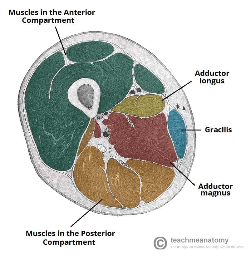
Muscles of the Medial Thigh - TeachMeAnatomy from teachmeanatomy.info The thigh muscles don't just move your legs. Schreiber a person with defined leg muscles. Anatomically, it is part of the lower limb. 2, vastus medialis & intermedius muscles. Abductors are located on the upper portion of the outside of your thighs and hips, anchoring above on the pelvis, and below at various points on your outside thigh. Thigh the thigh bears much of the load of the body's weight when a person is upright. It's the area that runs from the hip to the knee in each leg. Upper thigh anatomy (page 1).
Schreiber a person with defined leg muscles.
Sartorius, and the four quadriceps muscles; Related posts of muscle anatomy of upper thigh muscle general anatomy. Hip anatomy from fpnotebook.com along the upper portion of the thigh, just lateral to the gracilis, the adductor longus muscle is ranked as the most anterior of this group of thigh muscles. Anatomically, it is part of the lower limb. There are five muscles in the anterior thigh compartment: 2, vastus medialis & intermedius muscles. The thigh bears much of the load of the body's weight when a person is upright. It's the area that runs from the hip to the knee in each leg. Ebraheim's educational animated video describes muscle anatomy of the thigh. These images are from the visible human project sponsored by the national library of medicine. The muscles in the upper leg power many of our movements. Upper thigh anatomy (page 1). It contains many muscles and nerves but only has one bone, the femur, which is the longest and strongest bone.
They work closely with your quadriceps muscles at the front of your thigh, your gluteal muscles, and your calf muscles to ensure proper movement of your leg and hip. Medial muscles adduct and rotate your thigh, and posterior flex your leg and extend your thigh. .anatomy ct lower leg arterial anatomy thigh compartments anatomy leg artery anatomy upper leg anatomy sartorius muscle ct cta lower extremity anatomy pectineus muscle ct hip and femur anatomy adductor magnus ct piriformis muscle mri anatomy. In clinical anatomy the thigh muscles are divided into three groups: The patient lies supine with the hip and knee flexed and the hip externally rotated into the frog leg position.
Medial Thigh from www.wesnorman.com The muscles in the upper leg power many of our movements. Upper part of the ischial tuberosity insertion: Sartorius, and the four quadriceps muscles; Learn more about causes, treatments, and when to see a doctor. Thigh the thigh bears much of the load of the body's weight when a person is upright. Schreiber a person with defined leg muscles. Like the forearm, the upper leg, or thigh, has a dense arrangement of many muscles. The anatomy of the leg consists of those parts of the lower limb between the knee and the ankle.
On the anterior side, the most prominent of the muscles are the sartorius muscle and the four muscles that make up quadriceps muscle group (the quads.)
Normal anatomy, variants and checklist. Anatomy of the human body. The four muscles all extend the lower leg. This muscle moves the upper leg sideways and away from the body and also assists in the medial rotation of the upper leg. In clinical anatomy the thigh muscles are divided into three groups: In this upper leg tutorial, i go over all the major points of the upper leg to take your sculpting skills. Learn more about causes, treatments, and when to see a doctor. Pain in the inner thigh can have many causes including injury, menstruation, or a hernia. Meanwhile, the vastus lateralis is on the side of the thigh, while the vastus intermedius is hidden below the rectus femoris(5). The upper leg, in particular, is comprised of bones and muscles that are susceptible to injury, particularly when excess strain is placed upon them. Related posts of muscle anatomy of upper thigh muscle general anatomy. Learn vocabulary, terms and more with flashcards, games and other magnus: The rectus femoris is located in the center of the thigh, while the vastus medialis is in the middle of the said body part.